Bone Cross Sections - Tutorial 24 Variables For Tubular Bones Asp Msp And Bone Density Sauropod Vertebra Picture Of The Week : Related posts of cross section of human bone diagram human back muscles and bones.
byAdmin•
0
Bone Cross Sections - Tutorial 24 Variables For Tubular Bones Asp Msp And Bone Density Sauropod Vertebra Picture Of The Week : Related posts of cross section of human bone diagram human back muscles and bones.. As the names suggest compact bone looks compact and the spongy bone looks like sponges. This is a short tutorial using blender 2.8 that shows how to create a bone cross section and using images to create the textures. Compact bone cross section courtesy: They are obtained by taking imaginary slices perpendicular to the main axis of organs, vessels, nerves, bones, soft tissue, or even the entire human body. The compact bone is made up of osteon.
Related posts of cross section of human bone diagram human back muscles and bones. Each bone in your body is made up of three main types of bone material: From wikimedia commons, the free media repository. Cross sections of bones and growth plates will result in many more knife defects. Diagram with articular cartilage, marrow, spongy bone, medullary cavity, endosteum, diaphysis, and periosteum.
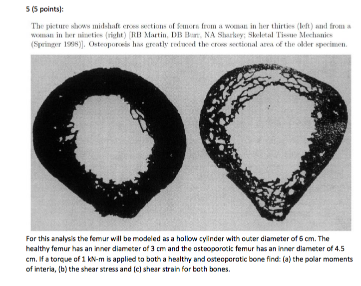
Solved 5 5 Points The Picture Shows Midshaft Cross Sec Chegg Com from d2vlcm61l7u1fs.cloudfront.net This is a short tutorial using blender 2.8 that shows how to create a bone cross section and using images to create the textures. This slide contained a cross section of a very small bone, and you are looking at the entire thickness of the shaft of the bone. Find the perfect bone cross section stock photos and editorial news pictures from getty images. Describes the bone markings, which are illustrated in (). The cortical bone equivalent area of the cross‐section of the region of interest (femoral neck or shaft), with all soft tissue voids (trabecular and cellular spaces) eliminated (cm 2). Download the photoshop bone brush here: There are three general classes of bone markings: This is known as the periosteum.
Describes the bone markings, which are illustrated in ().
Looking at a bone in cross section, there are several distinct. Related posts of cross section of human bone diagram human back muscles and bones. This is a short tutorial using blender 2.8 that shows how to create a bone cross section and using images to create the textures. Bones and tissues are studied by two different methods. Detailed and high textured 4k normal,disp,diffuse. Two types of bone tissues in cross section of a long bone : There are trabeculae in spongy bone which gives its sponge like appearance. The cortical bone equivalent area of the cross‐section of the region of interest (femoral neck or shaft), with all soft tissue voids (trabecular and cellular spaces) eliminated (cm 2). Translated into dutch.the original can be viewed here: In addition, cortical bone thickness at anterior, posterior, medial, and lateral parts of the bone section was measured. Create 2d cross section—if a 2d cross section does not exist in the drawing or if you want to. As the names suggest compact bone looks compact and the spongy bone looks like sponges. You may do so in any reasonable manner, but not in any way.
This slide contained a cross section of a very small bone, and you are looking at the entire thickness of the shaft of the bone. Compact bone is the outer layer and the spongy bone forms the inner layer. This is a retouched picture, which means that it has been digitally altered from its original version.modifications: That is, answer all cases of the question. Histology slide courtesy of william l.
Cross Section Bone High Resolution Stock Photography And Images Alamy from c8.alamy.com Photomechanical print page item number: A photoshop speed painting to show you how i quickly render out bone cross sections for medical illustration. A neutron can have many types of interactions with a nucleus. Click on a date/time to view the file as it appeared at that time. Looking at a bone in cross section, there are several distinct. Derivative works of this file: The cortical bone equivalent area of the cross‐section of the region of interest (femoral neck or shaft), with all soft tissue voids (trabecular and cellular spaces) eliminated (cm 2). In the last decade, considerable technological improvements have been made to repair damaged bones and tissue, such as bone cross sections with implants for microscopic examinations.
Cross‐sectional area is derived from the integral of the bone mass profile across the narrow region.
A neutron can have many types of interactions with a nucleus. It consists of two layers; Human bone cross section stock photos and images. The infobox for that structure appears on the left of the screen. Thin sections are much more common and provide considerably more information than bulk specimens and will be discussed in detail. An outer 'fibrous layer' containing mainly fibroblasts, and an inner 'cambium layer' containing progenitor cells. This is a short tutorial using blender 2.8 that shows how to create a bone cross section and using images to create the textures. Bones and tissues are studied by two different methods. Detailed and high textured 4k normal,disp,diffuse. (area/long bone length 3) ∗ 10 8. A photoshop speed painting to show you how i quickly render out bone cross sections for medical illustration. • a local cross section uses a breakout to see through an. Describes the bone markings, which are illustrated in ().
You may do so in any reasonable manner, but not in any way. • a local cross section uses a breakout to see through an. Browse 4,275 bone cross section stock photos and images available, or search for human bone cross section to find more great stock photos and pictures. Cross‐sectional area is derived from the integral of the bone mass profile across the narrow region. Translated into dutch.the original can be viewed here:
4 035 Bone Cross Section Illustrations Clip Art from media.istockphoto.com Photomechanical print page item number: Most relevant best selling latest uploads. A neutron can have many types of interactions with a nucleus. Cross section of bone diagram. Browse 53 bone marrow cross section stock photos and images available, or search for bone cross section or bone cells to find more great stock photos and pictures. In the last decade, considerable technological improvements have been made to repair damaged bones and tissue, such as bone cross sections with implants for microscopic examinations. Image.shutterstock.com if you look at the cross section of a long bone under a microscope, the rings of bone immediately internal to the periosteum of the bone are called _____. Diagram with articular cartilage, marrow, spongy bone, medullary cavity, endosteum, diaphysis, and periosteum.
Compact bone, spongy bone, and bone marrow.
Hope you enjoy and please. Thin sections are used for microradiography and for observation with transmitted light. A cross section of a human long bone. In the last decade, considerable technological improvements have been made to repair damaged bones and tissue, such as bone cross sections with implants for microscopic examinations. Related posts of cross section of a long bone bone test anatomy and physiology. • a local cross section uses a breakout to see through an. A photoshop speed painting to show you how i quickly render out bone cross sections for medical illustration. The cortical bone equivalent area of the cross‐section of the region of interest (femoral neck or shaft), with all soft tissue voids (trabecular and cellular spaces) eliminated (cm 2). Browse 53 bone marrow cross section stock photos and images available, or search for bone cross section or bone cells to find more great stock photos and pictures. There are trabeculae in spongy bone which gives its sponge like appearance. Human back muscles and bones 12 photos of the human back muscles and bones human back muscles and bones, bone, human back muscles and bones To the left is muscle tissue, and to the right is bone marrow. Describes the bone markings, which are illustrated in ().
Bone test anatomy and physiology 12 photos of the bone test anatomy and physiology anatomy and physiology bone lab test, anatomy and physiology bone markings test, anatomy and physiology bone practical test, anatomy and physiology bone tissue test, anatomy and physiology test on bone tissue, bone, anatomy and bone cross section. This slide contained a cross section of a very small bone, and you are looking at the entire thickness of the shaft of the bone.