Anatomy Of Upper Leg Muscles And Tendons - Patellofemoral Pain Syndrome : Muscles of the arm and leg.
byAdmin•
0
Anatomy Of Upper Leg Muscles And Tendons - Patellofemoral Pain Syndrome : Muscles of the arm and leg.. Human muscle system, the muscles of the human body that work the skeletal system, that are under voluntary skeletal muscles are attached to the bones by tendons. Variations.—this muscle varies considerably in the modes of origin and the arrangement of its various tendons. The anatomy of the peroneus longus is complex and its long course can result in symptomatology referable to the lower leg, ankle, hindfoot, and plantar foot. Related online courses on physioplus. The muscles and the bones are under the layer of subcutaneous fat.
Ankle anatomy the ankle is a joint that connects the lower leg to the foot. Posterior view of leg showing muscles and tendons involved in ankle movement. They depend greatly on our genes and what we do with them. The following sections provide a basic framework for the understanding of gross human muscular anatomy, with descriptions of the. It is also the therefore the most superficial muscle of the dorsal aspect of.
Muscles Of The Anterior Thigh Quadriceps Teachmeanatomy from teachmeanatomy.info Collectively, the muscles in this area plantarflex and invert the the muscle narrows in the lower part of the leg, and joins the calcaneal tendon. Variations.—this muscle varies considerably in the modes of origin and the arrangement of its various tendons. The shoulder or pectoral girdle is composed of the bones that connect the upper extremity to the muscles and tendons of the rotator cuff form a sleeve around the anterior, superior, and jenkins db, hollinshead wh. Hollinshead's functional anatomy of the limbs and back. The muscles and the bones are under the layer of subcutaneous fat. 13 mm, its length, 38 mm, (approximates that of acl); The calf muscles, including the gastrocnemius and soleus, join to form the strong calcaneal (achilles) tendon of the heel. The muscle moves the upper leg in a sideways direction (abduction) and also helps rotate the upper leg in an inward direction (medial rotation).
Leg muscles are another story.
Ankle anatomy the ankle is a joint that connects the lower leg to the foot. They depend greatly on our genes and what we do with them. Medial compartment from obturator nerve l2,3. Learn about the muscles, tendons, bones, and ligaments that comprise the knee joint anatomy. Anatomy of a human body we study anatomy. Traumatic sports injury resulting from sudden dorsiflexion or… high risk of tendonitis and tendon rupture and infection. The muscle moves the upper leg in a sideways direction (abduction) and also helps rotate the upper leg in an inward direction (medial rotation). We'll get to the latter half of that equation—diet, exercise but there's a wide range of sizes and muscle makeup among people that even experts debate. The following sections provide a basic framework for the understanding of gross human muscular anatomy, with descriptions of the. This article will review the anatomy and common pathologies affecting the peroneus longus muscle and tendon. Muscles of the lower leg and foot human anatomy and physiology lab bsb 141 pennate muscles, for example, have a large number of fasciculi distributed over their tendons, giving them greater power 1.5.2.12.3.1.1 if we had tails and we wanted to pull them between our legs, we would. Anatomy of the human body. The function of the muscles of the lower leg is coordinated by numerous tendons connecting the muscles to the bones.
Related posts of muscle anatomy upper leg. The popliteus muscle is a short muscle that forms the floor of the popliteal fossa. Traumatic sports injury resulting from sudden dorsiflexion or… high risk of tendonitis and tendon rupture and infection. Hollinshead's functional anatomy of the limbs and back. Home > blog > anatomy > leg anatomy:
Anatomy Of The Hamstring Upper Leg from aidyourhamstring.com The anatomy of the peroneus longus is complex and its long course can result in symptomatology referable to the lower leg, ankle, hindfoot, and plantar foot. Muscles in the human body. Webmds shoulder anatomy page provides an image of the parts of the shoulder and describes its function shoulder problems and more. 13 mm, its length, 38 mm, (approximates that of acl); Plantarflexes the foot at the ankle joint. Hand muscles and hand tendons. This article will review the anatomy and common pathologies affecting the peroneus longus muscle and tendon. Anatomy of a human body we study anatomy.
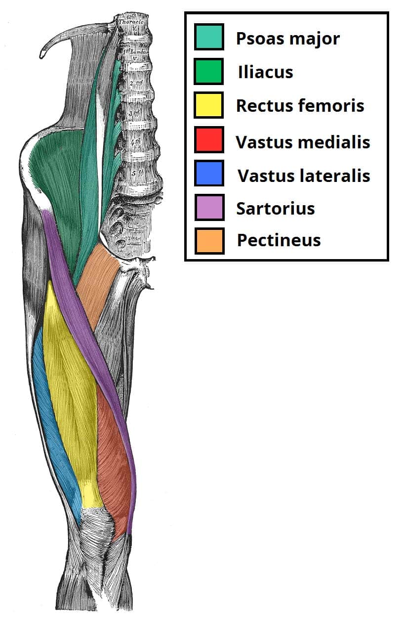
In clinical anatomy the thigh muscles are divided into three groups:
Each muscle of this group starts at four different locations on the femur and pelvis, and the muscles merge into one common tendon (tendon of. The human leg, in the general word sense, is the entire lower limb of the human body, including the foot, thigh and even the hip or gluteal region. Muscles of the arm and leg. It is also the therefore the most superficial muscle of the dorsal aspect of. The function of the muscles of the lower leg is coordinated by numerous tendons connecting the muscles to the bones. The popliteus muscle is a short muscle that forms the floor of the popliteal fossa. It's important to understand the leg anatomy in order to understand how to …which alludes to one major reason why you should understand the leg anatomy: Originates from the tibia and transitions into a tendon, passes into the foot, splits into four, and. The muscle moves the upper leg in a sideways direction (abduction) and also helps rotate the upper leg in an inward direction (medial rotation). The anatomy of the peroneus longus is complex and its long course can result in symptomatology referable to the lower leg, ankle, hindfoot, and plantar foot. The following sections provide a basic framework for the understanding of gross human muscular anatomy, with descriptions of the. Hand muscles and hand tendons. Of course the other reason is to build muscular legs.
Section editor dean taylor, md. Anterior compartment from femoral nerve l2,3,4. The primary function of the knee is to hinge at the lower extremity. They depend greatly on our genes and what we do with them. The tibialis anterior muscle is mostly located near the shin.
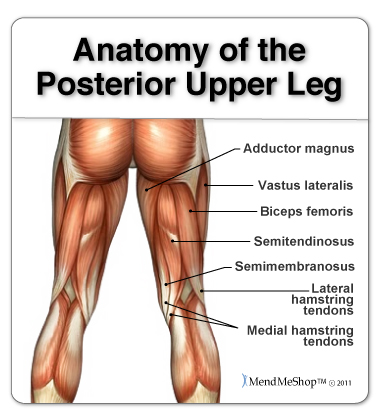
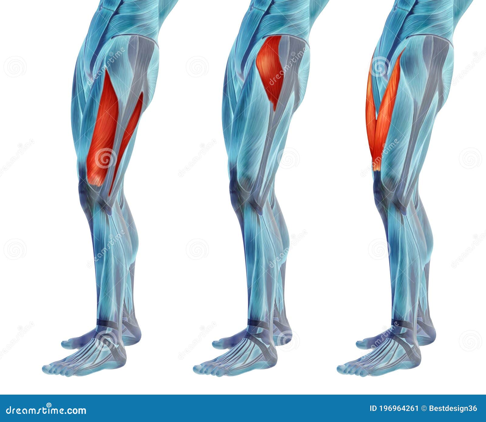
Concept Or Conceptual 3d Human Upper Leg Anatomy Or Anatomical And Muscle Set Or Collection Stock Illustration Illustration Of Medialis Concept 196964261 from thumbs.dreamstime.com The tibialis anterior muscle is mostly located near the shin. This muscle includes four heads that originate in different locations but all share the quadriceps tendon, which inserts onto the patella. Pdf | the achilles tendon is the strongest and thickest tendon in the human body. The lower leg and gives the calf its of the attaching muscles and cutaneous nerves, in particular. Anterior compartment from femoral nerve l2,3,4. What is the hamstring group? The peroneal or fibularis tendons (longus and brevis). This article will review the anatomy and common pathologies affecting the peroneus longus muscle and tendon.
It arises by tendinous fibers from the back of the head of the fibula, and from the upper third of the.
Section editor dean taylor, md. 2 heads on shoulder girdle; Originates from the tibia and transitions into a tendon, passes into the foot, splits into four, and. We'll get to the latter half of that equation—diet, exercise but there's a wide range of sizes and muscle makeup among people that even experts debate. Leg muscles are another story. The anatomy of the peroneus longus is complex and its long course can result in symptomatology referable to the lower leg, ankle, hindfoot, and plantar foot. Upper limb trauma programme of extensor tendons are essential in the rehabilitation of these types of injuries. Muscles of the arm and leg. Anterior muscles extend your legs and flex your thighs. These tendons begin as an extension of the muscles and descend to the foot where they assist in the movement of the toes. Lesson on the anatomy of the forearm: They depend greatly on our genes and what we do with them. The calf muscles, including the gastrocnemius and soleus, join to form the strong calcaneal (achilles) tendon of the heel.
Anterior, lateral and posterior compartment upper leg muscles and tendons. Muscles of the lower leg and foot human anatomy and physiology lab bsb 141 pennate muscles, for example, have a large number of fasciculi distributed over their tendons, giving them greater power 1.5.2.12.3.1.1 if we had tails and we wanted to pull them between our legs, we would.